Bubble contrast echocardiogram (echo)
Introduction
This leaflet has been written to give you information about your planned bubble contrast echocardiogram.
What is a bubble contrast echocardiogram?
You may have already had an echocardiogram performed. This is a non-invasive imaging test using ultrasound to look at your heart. Ultrasound is made of high-frequency sound waves which cannot be heard by the human ear. It is used to gain information regarding the structure and function of the heart muscles, chambers of the heart and structures within the heart such as the valves. The test involves placing a probe on the chest and does not use radioactivity.
A bubble contrast echocardiogram uses ultrasound combined with an injection of microbubbles to help determine additional information.
Why am I being asked to come for this test?
Your doctor is investigating the possibility you have a small hole or flap in the heart, located in the wall separating the left and right upper chambers of the heart (atria). Significant holes in your heart are usually detected in childhood. However, if there is a small flap or hole, this may not come to light until adulthood.
The microbubble contrast allows for the detection of these small holes as they do not usually show up on a normal echocardiogram.
Why might there be a defect in this part of my heart?
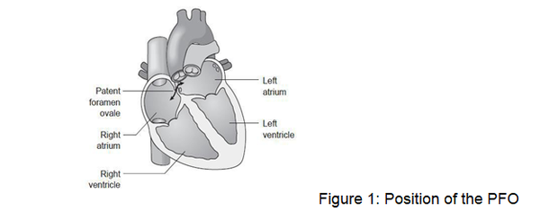
During the normal development of the foetal heart, there is an opening through the inter-atrial septum which allows blood to bypass the lungs which are not being used. Normally, this opening closes in the first few days or weeks after birth, but if it does not, the child will have a communication between the left and right atria.
This may take the form of a hole (Atrial Septal Defect – ASD) or flap (Patent Foramen Ovale – PFO) which behaves rather like a ‘trapdoor’. The defect will often correct itself without any medical intervention before the child reaches the age of 2, but about 25 to 30% of adults in the general population are said to have a PFO.
What happens if I have a PFO?
Many people do not have any symptoms or problems because of this defect, and it is detected by chance.
In some people, symptoms can result from blood clots forming in one of the veins in the leg (a deep vein thrombosis – DVT) and then a fragment of clot (embolus) passes from the right to the left side of the heart through the PFO; this then may block an artery resulting in:
- Stroke (loss of brain function)
- Heart attack (damage to the heart muscle)

Alternative reasons to perform the test may include:
- Extra communications (small holes) between blood vessels in your lungs (known as pulmonary arterio-venous malformations)
- Chronic liver disease
What does the echo involve?
- You will be taken into a room with usually a cardiac physiologist
- If you wish you may bring a friend or relative as an impartial observer (a ‘chaperone’) to be present during your examination
- You will be asked to undress to the waist and will be offered a hospital gown that should be left open to the front (like a coat). You will then be asked to lie on a couch. ECG stickers will be attached to your chest and connected to the echocardiogram machine. This will monitor your heart rate and rhythm during the test
- You will have a small plastic tube (cannula) inserted into one of the veins in your arm. This will be used later for the injection of microbubbles. You will then be asked to lie onto your left hand side. If you are unable to lie on your left side, we can carry out the echo while you are lying on your back. The test is performed in semi-darkness so the lights will be dimmed once you are comfortable
- The cardiac physiologist will place the echocardiogram probe on your chest and cold lubricating jelly (this helps to get good contact with the skin)
- If you have already had an echocardiogram, we will go straight on to perform the bubble contrast study. If not, several pictures of the heart will be recorded from different areas of your chest
- Once the baseline study has been completed, we will go on to the bubble contrast study. The bubbles are made up in a syringe using sterile saline (salty water) mixed with a little bit of air and a little bit of your blood drawn back from the vein via the cannula.
- These are rapidly mixed up to make very tiny microbubbles which are then injected into your vein. We will record pictures and watch carefully to see if any bubbles cross through from the right to the left side of the heart
- You will then be asked to cough and sniff, and with further injections you will need to perform a special breathing and blowing technique called the Valsalva manoeuvre; this will be carefully explained to you and practiced afterwards before we undertake this part of the test on the day
- The test will take about 45-60 minutes to complete
Do I need to take any special precautions before the test?
No, you should take all your usual medication as normal on the day of the test. You can also eat and drink normally. We advise that you keep hydrated (having plenty to drink) and keep yourself warm before the test. This increases the chance that we can access a vein for the cannula insertion during the test.
Is injecting air into the bloodstream harmful?
If a large amount of air was injected into a vein as a large bubble, it could potentially cause harm. However, the bubbles injected in this test are very small. If there is no hole in the inter- atrial septum, the bubbles will simply be filtered out by the lungs.
If you have a Patent Foramen Ovale (PFO) some bubbles will appear on the left side of the heart and then will gradually make their way through the circulation and be filtered out through the lungs.
Are there any risks in having the bubble contrast echo?
It is a very safe test. There are a very small number of reports world-wide of stroke or mini-stroke following this procedure, but the risk is very low. You may have some discomfort from the area where the cannula was inserted in your arm.
After the procedure
Once the test is complete you can get dressed and leave. There are no limitations to what you can do after the scan, for example, you may drive.
When will I receive the results?
The images are analysed on special software after the test and the result of this will be sent to your consultant. Your consultant will give you your results at your next clinic appointment.
Contact information:
If you have any questions about your planned dobutamine stress echocardiogram, please contact (Monday to Friday, 9:00am to 4:30pm):
Diagnostic Cardiology – ECG Department (Chelsea and Westminster Hospital)
- Tel: 020 33153443 / 5
Diagnostic Cardiology – ECG and ECHO Department (West Middlesex University Hospital)
- Tel: 020 8560 5336
This leaflet was written by:
Teresa Rutigliano (Senior Chief Cardiac Physiologist)
Reviewed and approved by:
Dr G Sunthar Kanaganayagam
Consultant Cardiologist and Cardiology Imaging Lead

